


Around the Eye
Your eyeball is surrounded by structures like the eyelids and the tear system. They protect, lubricate, and support the eye.
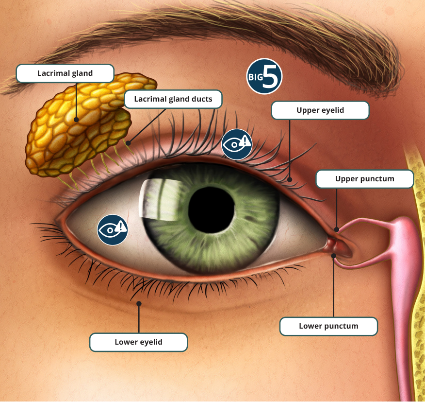
Lacrimal gland
The lacrimal gland is a small organ located above the eye. It produces lacrimal fluid (the watery layer of our tear film).
Lacrimal gland ducts
The Lacrimal gland ducts are drainage ducts that release the tear fluid onto the eye’s surface. As you blink, the tears are spread across the surface of the eye. Tears help to cleanse, lubricate, and nourish the eyes.
Upper eyelid
The eyelid is a fold of skin that closes over the eye to protect it.
Lower eyelid
The eyelid is a fold of skin that closes over the eye to protect it.
Upper punctum
The upper and lower puncta (singular punctum) are small openings on the upper and lower eyelids that drain tears from the eyes. The tears then travel through tear ducts into the back of the nose.
Lower punctum
The upper and lower puncta (singular punctum) are small openings on the upper and lower eyelids that drain tears from the eyes. The tears then travel through tear ducts into the back of the nose.
Styes and chalazia are both common bumps that form on or along the edge of the eyelid.
Stye (hordeolum)
A stye is a red, painful lump that grows from the base of an eyelash or on the inside of the eyelid. Most are caused by bacterial infection of a lash follicle or an oil-producing gland in the eyelid.
Chalazion
A chalazion is a swollen bump (or cyst) on the eyelid that occurs when an oil gland on the eyelid becomes clogged and inflamed. They can cause tenderness and can sometimes cause the entire eyelid to swell.
Learn more from expert Dr. Plemel.
Dry eye
Dry eye is a condition affecting the cornea caused either by not enough tears, or by poor quality tears. The result is a lack of lubrication in the eye, which can feel uncomfortable. Symptoms can include a gritty or stinging sensation, stringy mucus in and around the eyes, and blurry vision. Sometimes, as a response to the discomfort from dry eye, excess tearing can happen.
Eye injury
Eye injuries are common and cause anything from minor corneal scratches to vision loss or even blindness. They can affect any part of the eye or surrounding structures (like the eyelid or orbital bone), depending on the nature of the injury.
Keep learning about eye injury.
How your eye works
Believe it or not, your eye works a lot like a camera. Light rays enter the eye through the cornea, pupil, and lens. The cornea and lens work together to bend the light to focus it sharply onto the retina at the back of the eye. Cells in the retina turn light into electrical signals. These signals travel through the optic nerve from the retina to the brain, where the signal is interpreted as an image.
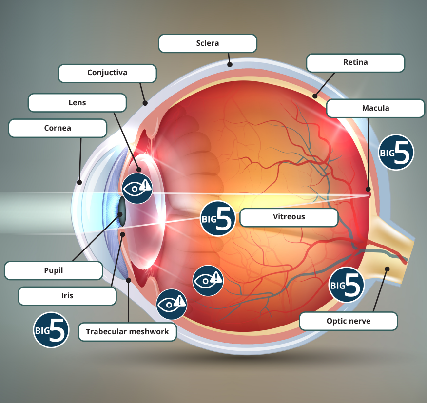
Cornea
The cornea is the clear, dome-shaped portion at the front of the eye. It helps focus light by bending it as it enters the front of the eye. In fact, the cornea is responsible for 70% of the eye’s focusing power.
Iris
The iris is the coloured part of the eye. The iris adjusts the size of the pupil to control the amount of light reaching the back of the eye. Muscles in the iris cause the pupil to constrict (become smaller) in bright light and dilate (become larger) in dim light.
Trabecular meshwork
The trabecular meshwork is the spongy tissue located in the eye’s drainage angle – the space where the the iris and the cornea meet. The trabecular meshwork acts as the eye’s drainage system. Most of the fluid produced inside the eye (called the aqueous humour) drains out of the eye via the trabecular meshwork and eventually makes its way back into the body’s circulatory system.
Pupil
The pupil is the opening in the middle of the iris that allows light to enter the eye.
Lens
The lens is a clear part of the eye that sits directly behind the iris and pupil. The lens’ job is to help focus light correctly onto the retina at the back of the eye.
Sclera
The sclera is the “white” of the eye. It’s a strong layer of protective tissue that covers most of the surface of the eyeball.
Conjunctiva
The conjunctiva is a thin, clear membrane that covers the sclera and lines the inner surface of your eyelids. It protects your eye from irritants and helps keep moisture in.
Retina
The retina is a layer of light-sensitive tissue that lines the back of the eye. It contains special cells called photoreceptors, which turn light into electric signals that travel to the brain through the optic nerve.
There are two types of photoreceptors: rods and cones. Rods perceive light levels, enabling vision in low light and at night. They’re concentrated in the outer areas of the retina and help give us our peripheral vision. Cones, which are concentrated at the centre of the retina, detect colour and provide central detailed vision.
Macula
The macula is a small area at the centre of the retina. It’s responsible for sharp central vision that allows us to clearly see details like faces and written text.
Vitreous
The vitreous (or vitreous humour) is a jelly-like substance. It fills the vitreous cavity, which is the space in the middle of the eye, between the lens and the back of the eye.
Optic nerve
The optic nerve is at the back of the eye and connects to your brain. It’s made of millions of nerve fibres that transmit electic impulses from the retina to the visual cortex – the part of our brain responsible for vision.
Retinal conditions
Two of the Big 5 serious eye conditions affect the retina.
Age-related macular degeneration (AMD)
AMD is a progressive condition that affects the macula, which is the small area at the centre of the retina that allows us to see fine details clearly. AMD happens when cells in the macula gradually begin to deterioriate, leading to loss of central vision.
Keep learning about AMD or hear from AMD expert Dr. Maberley.
Diabetic retinopathy
Diabetic retinopathy is an eye disease that commonly affects people with diabetes. Over time, high blood sugar levels caused by diabetes can damage blood vessels in the retina. There are various stages/types of diabetic retinopathy, but all will lead to vision loss if left untreated.
Keep learning about diabetic retinopathy.
Retinal tear
As we get older, the vitreous gel inside the eye changes to a more liquid consistency and separates from the retina, creating a posterior vitreous detachment (PVD). Usually, this happens without any issue. In rare cases however, as the vitreous separates from the retina, it pulls with enough force to cause the retina to tear. This can cause a sudden onset of floaters and/or light sensitivity. Retinal tears can lead to retinal detachment.
Retinal detachment
Retinal detachment happens when the retina is pulled away from the back wall of the eye. This can happen when the vitreous passes through a retinal tear and lifts the retina off the back of the eye.
Signs of a retinal detachment include the sudden onset of floaters, flashes, or a gray curtain moving across the field of vision. A retinal detachment is very serious and usually causes blindness if not treated urgently.
Glaucoma
Glaucoma is a disease characterized by damage to the optic nerve. This damage is usually related to an increase in pressure inside the eye.
The aqueous humour is a fluid that is produced inside the eye and drains out of the eye at the trabecular meshwork. If too much fluid is being produced, or if it is not draining properly (for example, if there is a problem with the trabecular meshwork), fluid builds up inside the eye, leading to a rise in pressure.
Click on the trabecular meshwork to learn more about it.
When eye pressure is too high, the nerve fibres of the optic nerve become damaged, which can cause blind spots and vision loss.
Keep learning about glaucoma or read our article featuring glaucoma expert Dr. Saheb.
Cataracts
As you age, the normal proteins in the eye’s lens begin to break down, causing the lens to become cloudy. This prevents light rays from passing clearly through the lens, resulting in hazy or blurry vision. When the lens becomes cloudy enough to obstruct vision to a large degree, it’s called a cataract.
Keep learning about cataracts.
Eye injury
Eye injuries are common and cause anything from minor corneal scratches to vision loss or even blindness. They can affect any part of the eye or surrounding structures (like the eyelid or orbital bone), depending on the nature of the injury.
Keep learning about eye injury.
Posterior Vitreous Detachment (PVD)
PVD is when the vitreous gel inside the eye pulls away from the retina. This happens because the vitreous becomes more liquid and shrinks slightly as we get older.
Many people with a PVD will not notice any symptoms, but some will notice a sudden increase in floaters and flashes in their vision. These are actually shadows cast on the retina by clumps of vitreous gel.
For most patients, these symptoms usually subside within 3 months of the PVD.
Rarely, PVD can result in serious complications like vitreous hemorrhage, retinal detachment, or macular hole. In these cases, flashes and floaters may be accompanied by decreased or distorted vision. This should be evaluated by an eye care professional immediately.
Conjunctivitis (“pink eye”)
Conjunctivitis refers to inflammation of the conjunctiva. It can caused by infection (either viral or bacterial) or by allergies. The small blood vessels in the conjunctiva become dilated, making your eyes look red. Symptoms can also include a burning or gritty sensation and a watery or sticky discharge.